
Diagnostic procedures
Various procedures are available for the diagnosis of cardiac diseases.
The electrocardiogram (ECG), cardiac ultrasound, blood analyses and stress tests are among the most frequently performed examinations.
Diagnostic procedures
Various procedures are available for the diagnosis of cardiac diseases.
The electrocardiogram (ECG), cardiac ultrasound, blood analyses and stress tests are among the most frequently performed examinations.
Non-invasive diagnostics
Non-invasive diagnostic procedures enable comprehensive cardiac analysis without invasive access to the body. These methods are particularly gentle on patients and provide valuable information for the early detection and assessment of heart disease.
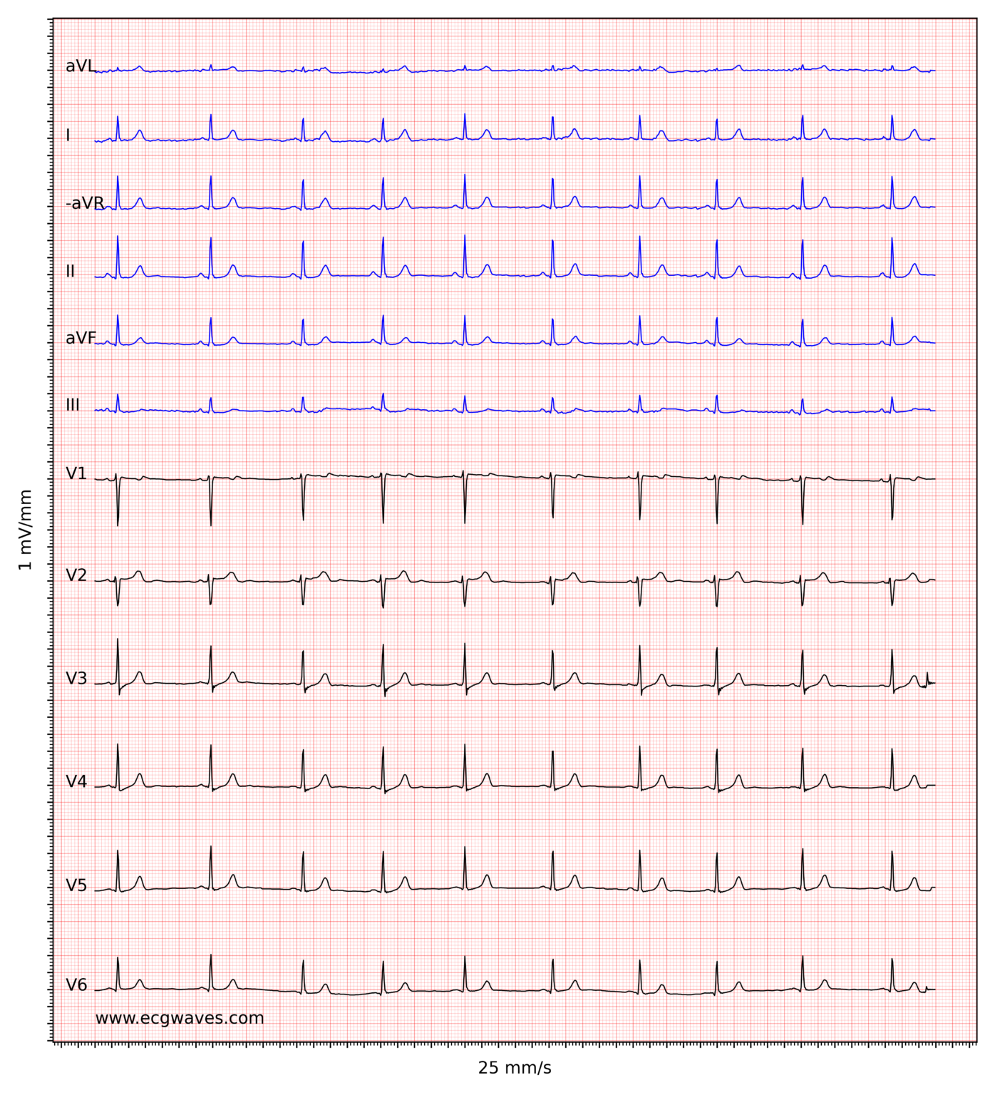 An electrocardiogram (ECG) is a simple and painless examination in which the electrical activity of the heart is measured. Small electrodes are attached to the skin, usually on the chest, arms and legs. The ECG shows how regularly and at what speed the heart beats and helps to detect cardiac arrhythmia or other heart problems at an early stage.
 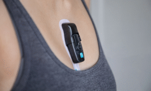 With the long-term ECG, the heart rate and heart rhythm are recorded for a maximum of 10 days. The extended recording period can make it possible to detect intermittent arrhythmias.
  Ergometry, also known as exercise ECG, measures physical performance while simultaneously monitoring various heart functions using an electrocardiogram (ECG).
 In spiroergometry, cardiac activity under stress is recorded in the same way as in ergometry. In addition, with the help of a well-fitting mask, the air inhaled and exhaled is also examined. This enables additional measurements such as maximum oxygen uptake.
 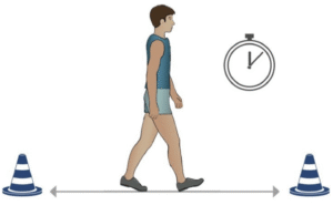 Another method of assessing patients’ resilience is to carry out a 6-minute walking test.
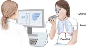 During the pulmonary function test, the lung and respiratory volume and the inhalation and exhalation rate are measured while the patient breathes into a spirometer via a mouthpiece.
 Taking a blood sample to assess central organ systems. Among other parameters, blood lipids (cholesterol), blood sugar levels and kidney function are checked.
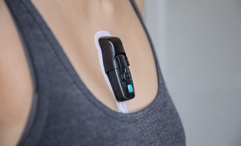 With the long-term ECG, the heart rate and heart rhythm are recorded for a maximum of 10 days. The extended recording period can make it possible to detect intermittent arrhythmias.
  Ergometry, also known as exercise ECG, measures physical performance while simultaneously monitoring various bodily functions using an electrocardiogram (ECG).
 During the pulmonary function test, the lung and respiratory volume and the inhalation and exhalation rate are measured while the patient breathes into a spirometer via a mouthpiece.
Cardiac Imaging
Imaging procedures provide a more precise insight into the structure and function of the heart. In addition to ultrasound examinations (TTE, TEE, 3D special examinations), computer tomography (CT), magnetic resonance imaging (MRI) and/or radionuclide procedures (SPECT/PET) provide important information on a variety of clinical pictures. Our doctors have proven expertise (EACVI Level III) and many years of experience in all cardiac imaging procedures.
 Ultrasound examination via the esophagus is used for a more detailed assessment of certain structures, in particular the heart valves.
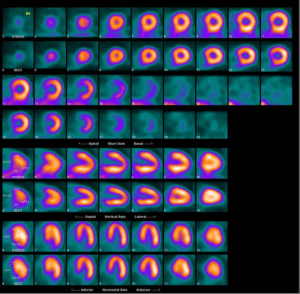 Radionuclide Imaging uses low-level radioactive “contrast agents” (so-called radionuclide tracers) to visualize structures, functions or even individual molecules in the heart. Scintigraphy is most commonly used to assess cardiac perfusion in cases of suspected
  Ultrasound examination via the esophagus is used for a more detailed assessment of certain structures, in particular the heart valves.
 The special procedure of cardiac scintigraphy is used for the imaging analysis of the blood flow and vitality of the heart muscle, for example in coronary heart disease.
Invasive cardiac diagnostics
In addition to non-invasive functional diagnostics and imaging procedures, our specialists also use invasive methods to examine and treat heart disease.
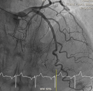 Coronary angiography (cardiac catheterization) is an imaging procedure used to visualize the coronary arteries in order to make stenosis or occlusions visible.
 Coronary angiography (cardiac catheterization) is an imaging procedure used to visualize the coronary arteries in order to make stenosis or occlusions visible.
Our specialists in cardiac diagnostics

Prof. Dr. med.
Prof. Dr. med.
Patric Biaggi
Patric Biaggi
Cardiology | Imaging
Cardiology | Imaging
DE – EN – FR – IT
DE – EN – FR – IT

Prof. Dr. med.
Prof. Dr. med.
Roberto Corti
Roberto Corti
Interventional cardiology
Interventional cardiology
DE – FR – IT – EN
DE – FR – IT – EN

Graduate doctor
Graduate doctor
Daniel Fritschi
Daniel Fritschi
Senior physician cardiology
Senior physician cardiology
DE – EN
DE – EN

Prof. Dr. med.
Prof. Dr. med.
Oliver Gämperli
Oliver Gämperli
Interventional cardiology
Interventional cardiology
DE – EN – FR – IT – ES
DE – EN – FR – IT – ES

Dr. med.
Dr. med.
Raffael Ghenzi
Raffael Ghenzi
Senior physician cardiology
Senior physician cardiology
DE – EN – FR – ES
DE – EN – FR – ES

PD Dr. med.
PD Dr. med.
David Hürlimann
David Hürlimann
Cardiology | Rhythmology
Cardiology | Rhythmology
DE – EN – FR
DE – EN – FR

Dr. med.
Dr. med.
Ioannis Kapos
Ioannis Kapos
Cardiology | Imaging
Cardiology | Imaging
DE – EN – GR
DE – EN – GR

Prof. Dr. med.
Prof. Dr. med.
Georg Noll
Georg Noll
Cardiology | Prevention
Cardiology | Prevention
DE – EN – FR – IT
DE – EN – FR – IT

Dr. med.
Dr. med.
Luca Oechslin
Luca Oechslin
Senior physician cardiology
Senior physician cardiology
DE – EN – IT
DE – EN – IT

Dr. med.
Dr. med.
Ivano Reho
Ivano Reho
Interventional cardiology
Interventional cardiology
DE – EN – IT – FR
DE – EN – IT – FR

Prof. Dr. med.
Prof. Dr. med.
Jan Steffel
Jan Steffel
Cardiology | Rhythmology
Cardiology | Rhythmology
DE – EN – FR
DE – EN – FR


