
Potential of fusion imaging and automated three-dimensional cardiac segmentation during transcatheter aortic valve replacement
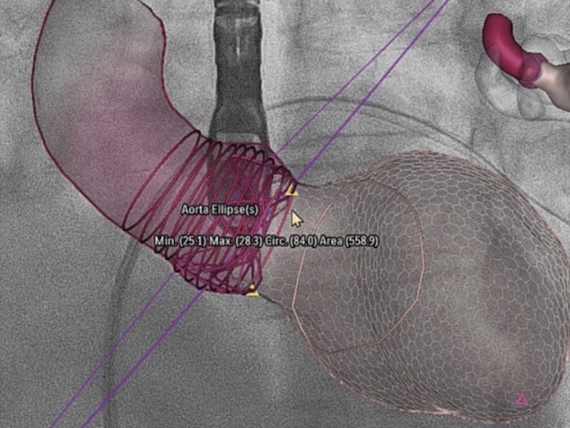
3D model of the aortic root and left ventricle with fluoroscopy.
Cardiac transcatheter aortic valve replacement (TAVR) is the preferred procedure for aortic valve replacement.
Pre-procedural multidetector computed tomography (MDCT) for aortic root analysis and valve prosthesis sizing is considered the standard of care.
However, the use of an iodinated contrast agent during MDCT angiography carries a significant risk of nephrotoxicity in patients with chronic severe renal failure. Three-dimensional (3D) transesophageal echocardiographic (TEE) imaging is considered a valuable alternative for aortic root sizing in this population.
– – –
(Published: December 2020)(PDF)
Patric Biaggi, Dominik F. Sager, Jeremy Külling, Silke Küest, Christophe Wyss, David Hürlimann, Ivano Reho, Ines Bühler, Georg Noll, Maurus Huber, Oliver Gaemperli, Peter M. Wenaweser, Roberto Corti
Source: Published on behalf of the American Society of Echocardiography. All rights reserved.